Venous Leg Ulcers, the 4Ds and Expected Healing Outcomes
What is a venous leg ulcer?
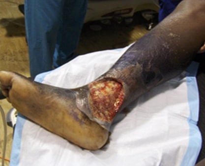
Venous leg ulcers are some of the most common lower extremity ulcers. They result as a late indicator of chronic venous insufficiency and venous hypertension. Normally, calf muscle contraction and intraluminal valves promote blood flow back to the heart while preventing blood reflux from gravity. However, when retrograde flow, obstruction, or both exist, the resultant chronic venous hypertension is responsible for potential damage to the venous system with resultant dermatologic and vascular complications that often result in the formation of a venous leg ulcer.

4 Ds For Healing Venous Leg Ulcers
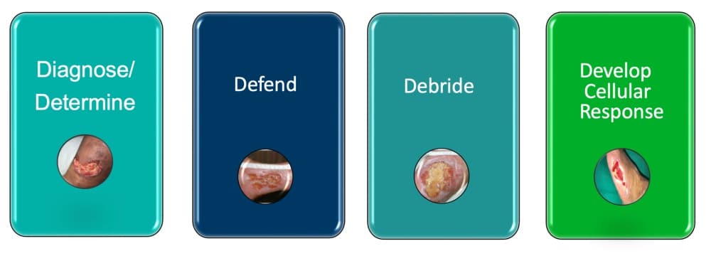
1. Diagnose and determine underlying factors impacting ulcer formation and healing
For all leg ulcers, a reliable diagnosis and evaluation of the underlying factors for non-healing are essential. The reliance on clinical examination with signs and symptoms of probable venous disease without supportive diagnostics may result in misdiagnosis or failure to identify and address the significant factors impacting healing. A study by Nelzén et al reported that a lack of systemic review and detailed testing, such as noninvasive arterial and venous Doppler and other diagnostics, results in misdiagnosis or treatment every fourth ulcer. It has also been reported that up to 20% of patients with venous leg ulcers have arterial compromise significant enough to delay wound healing.
2. Defend intact skin and wound tissue

Periwound skin care is perhaps most challenging in patients with venous leg ulcers. A number of physiological changes in the skin are caused by changes in the venous circulation in such patients. Patients with chronic venous ulcers often have lipodermatosclerosis, hemosiderosis, atrophy blanche, hyperpigmentation, dry, scaling and atrophic skin, and venous stasis eczema. There is also a common lymphatic system insufficiency which impacts the ability to drain interstitial fluid that accumulates in cases of severe chronic venous hypertension, further complicating edema and weeping tissues.
Peripheral edema of the lower extremity is typically present. It is detrimental to the lower extremity because it impacts the diffusion distance between blood capillaries and the tissues they supply. The tissues become deprived of adequate oxygen and nutrients, and metabolic waste products build up in an insufficient venous system. The periwound skin is vulnerable, thin, and easily damaged. Skin conditions can be further complicated by wound exudate and allergic or irritant reactions to topical therapies.
Bioburden of the skin and wound may also be problematic. Patients with venous leg ulcers are at risk for infection. Differentiating acute cellulitis, venous eczema, or acute lipodermatosclerosis is challenging as they all may present with erythema and edema, which often occur simultaneously.
Compression is the mainstay of management of venous leg ulcers, together with skin and local wound care. Leg elevation above the level of the heart during rest was recommended at one time, though shown not to be practical, and is not included in most current care practice. Newer treatments focus on careful selection of the right compression therapy and compression product to meet the individual needs of the extremity, the patient’s activity level, and the best option of compression type for patient adherence to the plan of care. In conjunction with compression, active calf muscle exercises should also be incorporated that are consistent with the individuals’ abilities. Appropriate weight loss, if indicated, and nutritional support and are also stressed.
3. Debride
Venous leg ulcers have highly proteinaceous exudate and tend to build up with fibrinous debris. This, in combination with a high bioburden and potential biofilm, makes debridement necessary. The goal is to address the aforementioned bioburden concerns – to remove slough, necrotic debris, and other nonviable items from the wound bed. This can be accomplished in a number of ways, dependent on the individual needs of the patient, wound, and site of care. It may include mechanical means such as monofilament pads, pulsed irrigation, or other debridement means such as autolytic, enzymatic, or surgical.
4. Develop a cellular response

Timely and effective interventions are needed to support a healing environment. A well-devised plan of care that incorporates the principles of advanced wound care must be implemented quickly and efficiently. This includes the early and appropriate assessment and differential diagnosis; an agreed-upon goal of care; management of bioburden and biofilm; protective and supportive skin care; effective compression therapy; and appropriate wound management, which often includes absorptive dressings, collagen, cellular- and tissue-based products, and grafts. Specific knowledge regarding skin care, including minimizing infection and proper application of advanced therapies, is essential to achieve proper healing.
Expected Healing Outcomes for Venous Leg Ulcers
Outcome factors that have been evaluated by Kanto and Margolis provide a prognostic index of healing in venous leg ulcers. Negative predictors they published included:
- Ulcer size ≥10cm²
- Duration ≥12 months
- History of venous ligation or venous stripping
- History of hip or knee replacement surgery
- ABI < 0.80
- Presence of fibrin on more than 50% of the wound surface
Data from the study also suggested that a venous leg ulcer that fails to decrease in size by 30% of its initial size over the first 4 weeks of treatment has a 68% probability to heal within 24 weeks. Recidivism rates for venous ulcers are high without proper management.
These findings underscore is the importance of timely care and the need to refer to a wound care specialist if the aforementioned predictors are identified.
To learn more about products that manage venous leg ulcers, please visit: https://sanaramedtech.com/surgical/
References:
O. Nelzén, D. Bergqvist, A. Lindhagen, Long-term prognosis for patients with chronic leg ulcers: a prospective cohort study, European Journal of Vascular and Endovascular Surgery, 1997:13(5):500-508.
Harding, Keith & C.Dowsett, & Fias, L. & Jelnes, Rolf & Mosti, Giovanni & Oien, Rut & Partsch, Hugo & Reeder, Suzan & Senet, P. & Verdú Soriano, José & Vanscheidt, Wolfgang. (2015). Simplifying Venous Leg Ulcer Management. Wounds International. Wounds International.
Kanto J. Margolis DJ. A multicenter study of percentage change in venous leg ulcer area as a prognostic index of healing at 24 weeks. Br J Dermatology. 2000:142(5):960-964.


